WikiJournal of Medicine/Ultrasonography of a cervical pregnancy
![]()
WikiJournal of Medicine
Open access • Publication charge free • Public peer review • Wikipedia-integrated
This article has been through public peer review.
Post-publication review comments or direct edits can be left at the version as it appears on Wikipedia.First submitted:
Accepted:
Reviewer comments
PDF: Download
DOI: 10.15347/wjm/2014.011
QID: Q44276856
XML: Download
Share article
![]() Email
|
Email
| ![]() Facebook
|
Facebook
| ![]() Twitter
|
Twitter
| ![]() LinkedIn
|
LinkedIn
| ![]() Mendeley
|
Mendeley
| ![]() ResearchGate
ResearchGate
Suggested citation format:
Jesper Agrell (27 November 2014). "Ultrasonography of a cervical pregnancy". WikiJournal of Medicine 1 (2). doi:10.15347/WJM/2014.011. Wikidata Q44276856. ISSN 2002-4436. https://upload.wikimedia.org/wikiversity/en/c/c4/Ultrasonography_of_a_cervical_pregnancy.pdf.
Citation metrics
AltMetrics
Page views on Wikipedia
Wikipedia: Content from this work is used in the following Wikipedia article: Cervical pregnancy.
License: ![]()
![]() This is an open access article distributed under the Creative Commons Attribution License, which permits unrestricted use, distribution, and reproduction, provided the original author and source are credited.
This is an open access article distributed under the Creative Commons Attribution License, which permits unrestricted use, distribution, and reproduction, provided the original author and source are credited.
Mikael Häggström ![]() (handling editor) contact
(handling editor) contact
Mikael Häggström ![]()
Article information
Abstract
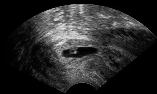
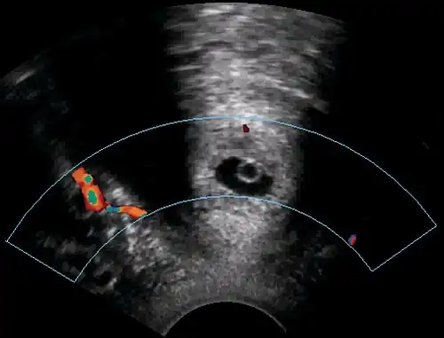
A woman in her 20s came to the gynecologic clinic because of a positive pregnancy test and a history of a previous ectopic pregnancy. The vaginal ultrasonography performed by Dr. Jesper Agrell showed a gestational sac in the cervix as displayed in the sagittal plane. The corpus of the uterus is located at right in the image. There was a discernible heartbeat, and the gestational sac diameter corresponded to a gestational age of 5 weeks. The distance from the gestational sac to the external orifice was only 15 millimeters.