6

I am attempting to measure the size of water in oil emulsions under a microscope. I generate the bubbles from a microfluidic device with a mineral oil continuous fluid ($n \approx 1.47$) and stabilized with a surfactant (1% w/w in oil). Inside the bubble is an aqueous solution of fluorescent protein.

When I image the bubble with bright field, there seems to be a dark band around the edge of the bubble, which I assume is due to total internal reflection. However, under the fluorescent channel, there appears to be a definite boundary which lies inside the dark band of the brightfield image.

Is the real size of the bubble the outside of the dark band in brightfield, or the boundary of the fluorescent image?

enter image description here

2 Answers2

3

The larger, dark outer shell is not indicative of the bubble size. However, the inner circle is.

It looks like the far left image is: (1) the fluorescent only channel, middle image: (2) brightfield, and right image: (3) fluorescent and brightfield.

It appears that in (1) the entire drop has a well-defined boundary/edge which corresponds to the drop-oil interface.

In (2) we now see a dark fringe around the drop which is larger than the boundary in (1). This dark ring is not a physical boundary, but is an artifact, primarily due to the fact that there is a refractive index mismatch$^1$ (with some possible diffraction), which causes the exaggerated edge and making the bubble look larger than it actually is.

In (3) we see the fluorescent boundary lies inside the dark ring in (2) and is of exact size compared to (1), thus confirming that the bubble (outer) edge in (2) is exaggerated.

Or simply put, the actual drop is the fluorescent image in (1) and the fluorescent image region of (3) which show the same physical drop.

$^1$ The bubble core/protein solution has a higher refractive index than the surrounding oil (you have also stated there's a surfactant you added, but these are typically thin enough not to cause significant refraction or total internal reflection). This means at the interface between bubble and oil, there is strong refraction. Also note that a quick google search shows that these oils are typically low refractive index fluorinated hydrocarbons with $n\approx 1.29$ - c.f., protein solution $\approx 1.33$.

joseph h
  • 31,821
0

The real size of the bubble is the outer edge of the dark band in the bright field image. Refraction of the fluorescent light exiting the bubble makes the fluorescent image smaller than the actual bubble.

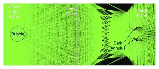
With some effort, this could be analyzed analytically. For example, Schwendener and Nägeli calculated the thickness of the dark annulus inside the edge of a air bubble in water to be $21$% of the radius of the bubble for a microscope aperture of $60$°. The diameter of the bright area inside the bubble was only 79% of the diameter of the actual bubble.

It is now perhaps easier to use simple ray tracing software to see the effect, e.g. a side view using the Ray Optics Simulation at https://phydemo.app/ray-optics/simulator/. A simple ideal lens focuses the image of the bubble. To make the effects larger and more visible, I have exaggerated the difference in index of refraction between outside and inside the bubble, i.e.

$$\frac{n_\mathrm{outside}}{n_\mathrm{inside}}=2$$

A backlit bubble produces a bright centre with a dark annulus. (Click here to access an editable version of the simulation.) Ray tracing for diffuse lighting of a low index bubble in an high index liquid

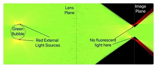
A fluorescent bubble produces an image smaller than the actual bubble. (Click here to access an editable version of the simulation below.) To make the apparent smaller size obvious, I added two red light sources just on the outside of the bubble, and the image shows the gap in the image between the apparent fluorescent image of the bubble and the red light from the actual outer edge of the bubble. Ray tracing of a low index fluorescent bubble in an high index liquid

To better see what is happening to fluorescent light near the edge of the bubble, here are just one of the external light sources plus a few selected fluorescent rays. The edge fluorescent light bends when leaving the bubble and ends up being focused to a smaller diameter image. A few selected fluorescent rays

David Bailey
  • 13,658