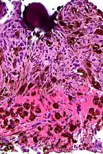
Micrograph of psammoma body in the centre of the field in a meningioma of brain. H&E stain.
A psammoma body is a round collection of calcium, seen microscopically. The term is derived from the Greek word ψάμμος (psámmos), meaning "sand".
Cause
Psammoma bodies are associated with the papillary (nipple-like) histomorphology and are thought to arise from,
- Infarction and calcification of papillae tips.
- Calcification of intralymphatic tumor thrombi.[1]
Association with lesions
Psammoma bodies are commonly seen in certain tumors such as:
- Papillary thyroid carcinoma[2]
- Papillary renal cell carcinoma[3]
- Ovarian papillary serous cystadenoma and cystadenocarcinoma[4]
- Endometrial adenocarcinomas (Papillary serous carcinoma ~3%-4%)
- Meningiomas, in the central nervous system[5]
- Peritoneal and Pleural Mesothelioma
- Somatostatinoma (pancreas)[6]
- Prolactinoma of the pituitary [7]
- Glucagonoma[8]
- Micropapillary subtype of Lung Adenocarcinoma[9]
Benign lesions
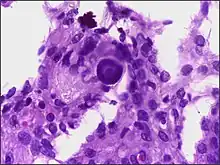
Micrograph of a psammomatous melanotic schwannoma with a psammoma body, as may be seen in Carney complex. H&E stain.
Psammoma bodies may be seen in:
- Endosalpingiosis[10]
- Psammomatous melanotic schwannoma
- Melanocytic nevus[11]
Appearance
Psammoma bodies usually have a laminar appearance, are circular, acellular and basophilic.
References
- ↑ Johannessen JV, Sobrinho-Simões M (September 1980). "The origin and significance of thyroid psammoma bodies". Lab. Invest. 43 (3): 287–96. PMID 7401638.
- ↑ Chapter 20 in: Mitchell, Richard Sheppard; Kumar, Vinay; Abbas, Abul K; Fausto, Nelson (2007). Robbins Basic Pathology. Philadelphia: Saunders. ISBN 978-1-4160-2973-1. 8th edition.
- ↑ "Renal Cell Carcinoma". The Lecturio Medical Concept Library. Retrieved 1 October 2021.
- ↑ Ovarian papillary serous cystadenocarcinoma at WebPath, The Internet Pathology Laboratory for Medical Education at Mercer University School of Medicine. Retrieved July 2011
- ↑ "Brain Stem & Posterior Fossa". Archived from the original on 2000-03-01.
- ↑ Lewis RB (2010). "Pancreatic Endocrine Tumors: Radiologic-Clinicopathologic Correlation". RadioGraphics. 30 (6): 1445–1464. doi:10.1148/rg.306105523. PMID 21071369.
- ↑ Robbin's Pathology, Eight Ed
- ↑ "Glucagonoma". The Lecturio Medical Concept Library. Retrieved 1 October 2021.
- ↑ Emoto K, Eguchi T, Tan KS, Takahashi Y, Aly RG, Rekhtman N, Travis WD, Adusumilli PS (2019). "Expansion of the Concept of Micropapillary Adenocarcinoma to Include a Newly Recognized Filigree Pattern as Well as the Classical Pattern Based on 1468 Stage I Lung Adenocarcinomas". J Thorac Oncol. 14 (11): 1948–1961. doi:10.1016/j.jtho.2019.07.008. PMC 8785415. PMID 31352072.
{{cite journal}}: CS1 maint: multiple names: authors list (link) - ↑ Hallman KB, Nahhas WA, Connelly PJ (September 1991). "Endosalpingiosis as a source of psammoma bodies in a Papanicolaou smear. A case report". J Reprod Med. 36 (9): 675–8. PMID 1774734.
- ↑ Rapini, Ronald. Practical Dermatopathology. Elsevier Mosby, 2005, p. 10.
External links
Look up papillary in Wiktionary, the free dictionary.
Slides:
This article is issued from Wikipedia. The text is licensed under Creative Commons - Attribution - Sharealike. Additional terms may apply for the media files.